Deep Learning-Based Segmentation of Peritoneal Cancer Index Regions from CT Imaging
arXiv cs.CV / 5/1/2026
📰 NewsDeveloper Stack & InfrastructureModels & Research
Key Points
- The paper addresses the gap between the invasive Sugarbaker’s Peritoneal Cancer Index (sPCI) assessment and the need for a standardized non-invasive imaging-based counterpart (radiological PCI, rPCI) defined using 3D regions.
- It proposes a deep learning method to automatically segment rPCI regions on CT, focusing on 13 anatomical regions to support imaging-based scoring.
- The study evaluates nnU-Net and Swin UNETR on 62 CT scans with rPCI manually annotated by three clinical researchers and validated by two expert radiologists.
- nnU-Net achieved an overall Dice score of 0.82, nearing interobserver agreement (0.88) and outperforming Swin UNETR (0.76), with most remaining errors concentrated in right flank and small-bowel regions.
- The results indicate that automated rPCI segmentation is feasible and could enable future non-invasive, imaging-based PCI assessment workflows.
Related Articles

Every handle invocation on BizNode gets a WFID — a universal transaction reference for accountability. Full audit trail,...
Dev.to
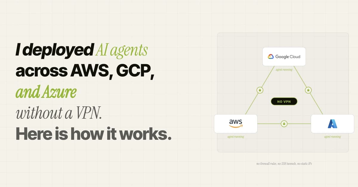
I deployed AI agents across AWS, GCP, and Azure without a VPN. Here is how it works.
Dev.to
Panduan Lengkap TestSprite MCP Server — Dokumentasi Getting Started dalam Bahasa Indonesia
Dev.to

Every Telegram conversation becomes a qualified lead. BizNode captures name, email, and business details automatically while...
Dev.to

MCP, Skills, AI Agents, and New Models: The New Stack for Software Development
Dev.to